Wednesday, March 23, 2016

Performance Enhancers and Creatine Satirical Ad


In the Performance Enhancement notes, we learned about different types of stimulants that can help people preform better and more efficiently.  People take these performance enhancers for many reasons, be it to aid in athletic ability, build muscle quicker, lose body fat faster, or even to help them recover faster. However, many of them can cause more damage than good. In both females and males, it can cause liver damage, high blood pressure, increased risk of tendon and ligament damage, and heart disease.

In males, it can cause the estrogen to come more into play. For example, testicles can shrink and their breasts can grow. In females, the opposite can occur. Testosterone levels will go up causing male-type facial and hair growth, deepening of the voice, and breast shrinkage.

There are many types of these stimulants that are well known such as caffeine, massages, and carbo-loading. These are the last risky stimulants. People (me included!) drink coffee every morning to get them to wake up, and this causes them to also be more alert and ready.

Other stimulants such as Erythropoietin(can cause stroke or heart attack), Anabolic steroids (can cause high blood pressure and liver damage), and Creatine (side affects listed in the above picture), are a lot more dangerous to use.

Thursday, March 17, 2016

Chicken Dissection Lab Analysis

In this lab, we used whole chickens from Safeway. We were instructed to find many different muscles by cutting into the back, front, wings and legs. In the first incision we cut along the midline of the chest to expose the pectoralis major and the pectoralis minor. These muscles help the bird move its wings, while in humans it pulls the shoulders forwards and down. These two muscles have striations running in opposite ways.

After this, we flipped the bird carcass onto its chest and did the same cut on the midline of the back. We peeled back the skin to expose the Trapezius and Latissimus Dorsi muscles. In humans, the trapezius either shrugs the shoulders, or pulls the shoulders back. The Latissimus Dorsi helps to extend the arm or wing.


We extended the wing of the bird next to get a better angle at the Deltoid, Biceps Brachii, and Triceps Humeralis. The Deltoid is located at the top of the shoulder to help with raising your arms. The Biceps Brachii flexes, or bends, the arm, while the Triceps Humeralis extends the arms. The second half of the wing, also our forearm, contained the Flexor Carpi Ulnaris, which flexes the hand, and the Brachioradialis, which pulls the hand back.

Moving onto the Chicken's legs, we found the Sartorius, Iliotibialis, Biceps Femoris, Semimembranosus, and Semitendinosus on the thigh area. The Sartorius flexes the thigh and is what allows humans to cross their legs, the Iliotibialis extends the thigh as well as flexes the knee. The Biceps Femoris flexes the leg, similarly to what the Biceps Brachii does in the arm. The Semimembranosus and Semitendinosus both help with extending the thigh.


The last section that we cut into was the drumstick, or the calf of humans. In this area, we found the Gastrocnemius, the Peroneus Longus, and the Tibialis Anterior. The Gastrocnemius extends the foot and flexes the lower leg, the Peroneus Longus also extends the foot, and the Tibialis Anterior flexes the foot.

After we did all of this, we started the cleanup process and disinfected the table, tools and our hands and forearms


Monday, March 14, 2016

What Happens When You Stretch


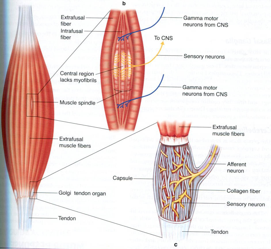
"When the muscle is stretched, so is the muscle spindle (aka intrafusal fibers). The muscle spindle records the length (and how fast) and sends signals to the spine which convey this information. This triggers the stretch reflex, or myotatic reflex, which attempts to resist the muscle length by causing the stretched muscle to contract. This basic function of the muscle fiber helps to maintain muscle tone and to protect the body from injury"
--I thought this quote was interesting because it says that the signals are sent to the spine. I'm not really sure why it is sent there. One of the reasons why we hold a stretch for a long time is because we need the muscle spindle to become accustomed to the new length so it stops sending signals.

"The static component of the stretch reflex persists as long as the muscle is being stretched. The dynamic component of the stretch reflex (which can be very powerful) lasts only for a moment and is in response to the initial sudden increase in muscle length."
-The static component is also known as the nuclear chain fibers, and the nuclear bad fibers are responsible for the dynamic component. Both of these are the two kinds of intrafusal muscle fibers.

"When muscles contract (possibly due to the stretch reflex), they produce tension at the point where the muscle is connected to the tendon, where the Golgi tendon is located."
- This is when the lengthening reaction occurs if the tension passes a certain point. The Golgi tendon is responsible for recording the tension.


Relate and Review:
Stretching starts when the sarcomere contracts which causes the areas between the thick and thin myofilaments to increase. When it stretches, the area decreases , and then the muscle fiber become longer. When you stretch the sarcomeres to their capacity, the connective tissue will then be able to stretch just a little bit further.


Tuesday, March 1, 2016

Unit 6 Reflection


    In this unit, although we have not done any blogging, we have learned a lot about the skeletal system. The skeleton is used for many different purposes. First, it supports the body. It also protects the bodies soft organs. The skeletal system not only includes 206 bones (luckily we only had to know and remember where 40 of them were), but it also includes cartilage, joints, ligaments(connects bone to bone), and tendons (connects bone to muscle).

    There are many different types of bones: long, short, flat and irregular. Long bones tend to be longer than they are wide and contain mostly compact bone. Short bones are typically cube-shaped such as carpals and tarsals. Flat bones are thin, flattened, and usually curved. Some examples of this are the sternum, skull, and ribs. The last type of bone is the irregular bone which does not fit into any other classification. This includes the vertebrae and hips bones.

    We also learned about 6 different diseases related to the skeletal system. Arthritis is a common disease where there are over 100 types. This is a disease where there is inflammation in the joints. Osteoporosis is when the bones have lost important minerals such as calcium, and then become brittle and porous. When bones are Vitamin D deficient, it cause softening and weakening of the bones, called Rickets. Scoliosis, Lordosis, and Kyphosis all have to do with the spine. Scoliosis is another commonly known disease that causes abnormal curvature of the spine side-to-side. When there is excessive curvature of the thoracic, or upper region of the spine, it is known as Kyphosis. Similarly, if there is excessive curvature of the Lumbar vertebrae, or lower region of the spine, it is known as Lordosis.


    I mentioned before that calcium was one of the main causes of Osteoporosis. Well, calcium actually aids a lot in health. It is necessary for maintaining pH of blood, heart function, nerve function, and the building and repairing of teeth and bones. It is very important to have healthy levels of calcium in your body, otherwise it gets pulled from our bones.
    In bone remodeling, there are 4 major cells that contribute. First, are the osteoprogenitor cells. They are dividing cells that develop into osteoblasts, which are the bone-building cells. Osteoblasts secrete collagen and osteocalcin. They then go on to become mature bone cells, or Osteocytes, which carry out the daily activities of bone tissue. Finally, there are Osteoclasts, which are found on the surface of the bones and are responsible for the breaking down of bone tissue.


    Another topic that we were taught was on bone fractures. We learned the difference between complete fractures (bone breaks completely through), incomplete or greenstick fracture (fracture does not extend through the bone), and closed or simple fracture (bone does not tear through the skin). It can get more complicated than this when it comes to factures, but I will leave it at this!


    I hope you enjoyed reading about my experience of learning about the human skeleton as much as I enjoyed learning about it! Thank You!

Wednesday, February 24, 2016

Owl Pellet Lab


Owl Pellet
Skull Top View
 In this lab, we were given an owl pellet to dissect. An owl pellet is something that an owl, usually a barn owl, regurgitates because they can't digest it properly. They usually regurgitate these pellets 2-3 times a day. The pellets mostly consist of fur and bones, so it does not break down. The possible organisms that you will be able to find are shrews, voles, rats, mice and birds.


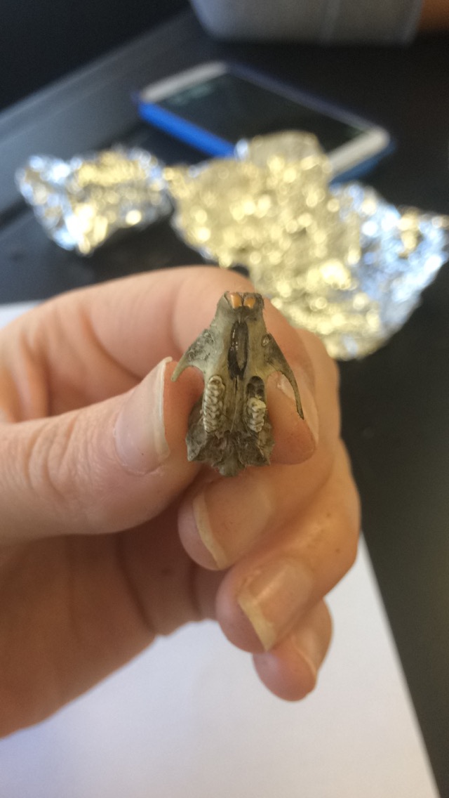
The owl pellet that we dissected ended up having a vole skeleton. Moles and voles are very similar, but we felt that the skeleton was a vole considering the scapula had a more defining corner instead of a curve. Although the pellet was only 4.7 cm long and 2.6 cm wide, we were able to find many bones. The skull and mandible that was produced were shaped like a voles, as well as many of the other bones that we extracted such as the forearm and scapula. It also consisted of many ribs, vertebrae, and tiny bones that were most likely phalanges or carpals. In all, our pellet weighed 6.62 grams.  
Skull Bottom View

As you can see in the picture of the whole skeleton put together, there are many bones that are similar to that of a human. First, the scapula's (aka shoulder blades) look very similar. Also, the vertebrae are categorized by the lumbar, thoracic, and cervical vertebrae. The Lumbar vertebrae are larger than the other two, just like with that of the human vertebrae. You can also see that the vole has a hip socket (black circular hole medial to the spine) where the femur will rotate.

Although, I oriented the bones like that of a human skeleton, the vole's skeleton should be oriented more on its side so that the legs and arms are overlapping. Or course, that would make the skeleton more confusing to look at. In taking a look at the skull of the vole, you can spot another difference. The eye sockets are located on the sides, whereas on a human, they are located in the front.  

This lab was both fun and difficult. It was fun extricating the bones from the pellet, although they were very fragile. It was difficult to piece them together since many of them were broken, as well as discovering what kind of organism it was. The bones were very similar to each other. I enjoyed this lab, and I hope you enjoyed reading about my experience!

Thank You!
Mole Skeleton
Human Skeleton

Thursday, January 28, 2016

Unit 5 Reflection

 "Unit 5 Reflection"


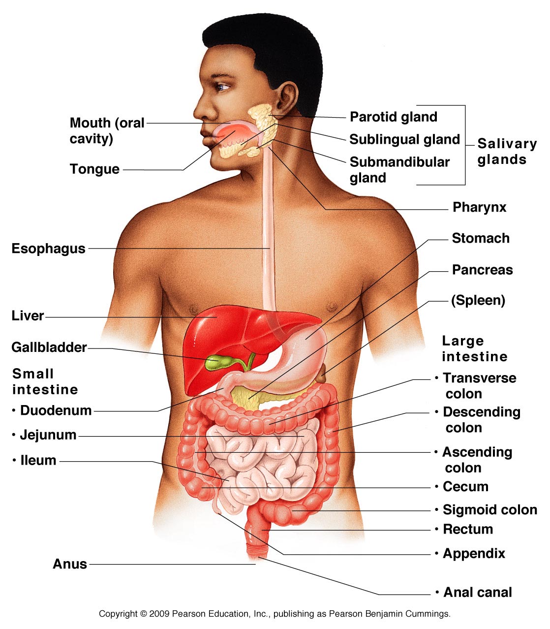
This unit was about the Digestive System, Metabolism, Endocrine System, and Lymphatic System. The Digestive System includes the Alimentary Canal which is the pathway that the food goes from your mouth to being defecated. The picture to the left shows all of the organs in the Digestive/Alimentary Canal. The digestive system is responsible for absorbing, digesting, secreting, and defecating. We did one lab for this unit about the digestive system which I had a previous blogpost as well and that can be found here. In our metabolism there are three stages. Stage 1 breaks down large molecules to smaller ones. Stage 2 takes those small molecules and turns them into Acetyl CoA. Stage 3 has common pathways such as the Krebs cycle and ETC that are oxidation for the fuel molecules. Also, there are three different states.  The Fed state is right after you eat. The pancreas releases insulin and the majority of glucose is taken by the liver to be converted into glycogen. The fasting state is where the insulin levels drop and the glucagon levels rise. The body also uses more fats for energy in order to conserve the glucose. The last state is the Starvation state. This occurs 4-5 days after eating where the muscles rely on adipose tissues for fuel. The Endocrine System controls the processes involved in movement and physiological equilibrium. There are many different glands such as the pituitary gland, the thyroid gland, and the parathyroid gland that are involved in this system. Some organs that are in the endocrine system are the pancreas, which secretes insulin or glucagon; gonads, which are either testes or ovaries that deal with testosterone or estrogen; and the kidneys, which regulate red blood cell production. The last system that we learned about in this unit was the Lymphatic System which specializes in immunity, lipid absorption, and fluid recovery. There are three different kinds of lymphocytes. T-cells, which you ca read more about here from my previous posts, deal with cellular immunity. Then there are b-cells for humoral immunity, and Natural Killer cells which provides immunological surveillance.

I think that this unit was very fascinating but it was a little bit difficult for me to keep up with all the new information. I really enjoy human anatomy and learning about the body, but when there is so much information to learn in a relatively short amount of time, it can be slightly daunting.


Thank you for reading! I hope you enjoyed it!

Amanda

Wednesday, January 6, 2016

Digestive System Lab


Digestive Organ
Length (cm)
Mouth
9.7 cm
Esophagus
42 cm
Stomach
19 cm
Small Intestine
721.36 cm
Large Intestine
180.34 cm
TOTAL
972.4 cm


For this activity, we measured the approximate lengths of each of the organs in our digestive system. For the mouth, we measured from our jaw to the front of our mouth. the esophagus was measured by going from our jaw to the end of our rib cage. The stomach was approximated by taking the length of the space between the thumb and pinky when the hand was stretched out. For the small intestine, we had to convert our height into centimeters and then multiply by 4. Lastly, the large intestine length was found my just taking our height in centimeters. This was an interesting way to figure out how long each organ was. The total length of my digestive system is almost 5 1/2 times the size of my height. This is a serious difference and it is amazing to think that all it would be able to fit in my body just by wrapping and coiling around. I would guess that, on average, it would take about 18 hours for food to move through my entire digestive system. In looking it up, the actual time is 24-72 hours. This makes sense considering people don't defecate every day, as my prediction would suggest. Some factors that may influence the time that it takes to pass through the whole digestive system are that the stomach needs time to break down the food, the bolus has to wind around 9 meters of organs, and in the process, many nutrients and other things are extracted from it on the way. Many muscles are used in this process to make the food used to its full potential. Digestion is different from absorption because digestion is the process of breaking down the food whereas absorption is taking in or soaking up substances or energy. The stomach and the small intestine are involved in the process of digestion, and the large intestine is involved mainly with the absorption aspect. The small intestine does help with absorption as well as digestion.